NOTES NURSING & MEDICAL STUDENTS Notes for medical student and nursing student
Lung:-
introduction :-
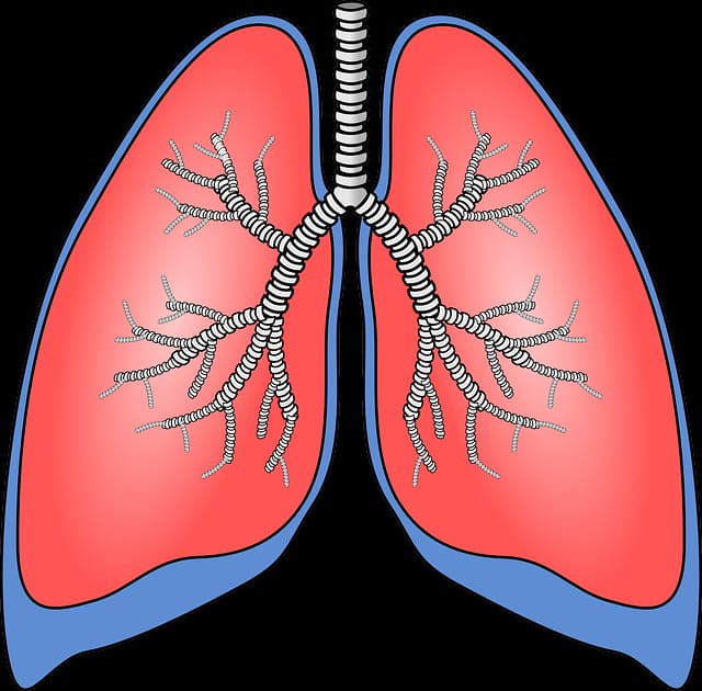
- lungs are a pair of respiratory organs situated in the thoracic cavity .
-the each lung innervates the corresponding pleural cavity .
- the lung young are brown or grey in color.
- they becomes mottled black because of the depositions of inhaled carbon particles .
relations of anterior part :-
!Right side :-
1) right atrium
2) small part of RV
3)SVC
4)right brachiocephalic
5) right vagus nerve
6) right phrenic nerve
7) trachea
!Left side :-
1) left ventricle
2)pulmonary trunk
3) arch of aorta
4) descending thoracic aorta
5) left vagus nerve
6)left recurrent
7)laryngeal nerve
Gross anatomy of lung :-
-there are a pair of lung in the thoracic cavity .
-there are 3 lobes in the right lung and 2 lobes in on left lung .
-there are two fissures in right lung , oblique and horizontal fissures .
-left lung has only one oblique fissures.
-each lung is covered by pleura base .protected by thoracic skeleton .
-each lung has an apex ,a broad base or infrior surface ,sharp anterior border ,and sharp inferior border .
- each lung has sterno costal surface , diaphragmatic surface and mediastinal surface.
-right lung is shorter than left due to right lobe of liver pushing the right lung superiorly .
Borders :-
1) Anterior border :-
-corresponds to the anterior " costomediastinal" line of pleural reflection .
-it is deeply notched in the left lung posterior to 5 th costal cartilage by the pericardium extends vertically downwards to from lingula .
2) Inferior border:-
-thin and shaep
-it separate the base of lung from the costal surface and extends into phrenicocotal sinus .
3) posterior border :-
-thick and ill defined .
-fits into deep paravertebral gutter
-extends from C7 to T10 .
Surfaces of lung :-
1) costal surfaces :-
-it is in contact with the costal pleura and overlying thoracic wall.
2) medial surfaces :- posterior , ventebral part ,anterior ,mediastinal part.
Horizontal fessure :-
- it extends from anterior margin at the level of the 4 th costal cartilage .
- runs horizontally backwards to meet the oblique fissure in the mid - axillary line .
-pulmonary pleura extends into the fissure of lungs so that the lobes can move on each other during respiration .
fissures and lobes of the lungs:-
- the oblique fissure cuts into whole thickness of the lungs, except at the hilum.
-due to the oblique plane the fisssure , the lower lobe is more posterior and the upper and middle lobe more anterior .
- in the right lungs the horizontal fissure passes from the anterior border upto the oblique fissure and separates a wedge shaped middle lobe from the upper lobe .
- the tongue shaped projection of the left lung below the cardiac notch si called lingula.
- the presence of the oblique fissure of the each lung allow a more uniform expantion of the whole lungs .
Broncho pulmonary segments :-
1) these are well defined sectors of the lung each of which is aerated by a tertiary or segmental bronchus .
2) each segment is pyramidal in shape with its apex directed toward the root of the lung .
3) these bronchopulmonary segments are independent respiratory units.
Blood supply of lungs :-
1) Arterial supply :-
- the right side there is on bronchial artery which arises from either the third posterior intercotal artery or from the upper left bronchial artery.
-the left side there are two brochial arteries both of which arises from the decending thoracic aorta .
-there are pre capillary anastomoses between bronchial and pulmonary arteries .
2) vagus drainage :-
-the usually are two bronchial veins on each side ,the right bronchial vein drain into the azygous vein.
-the left bronchial vein drain either into the left superior intercostal vein or into hemi azygous vein.
-the greater part of the venous blood from the lung is drained by the pulmonary vein.
3) Nerve supply :-
1) para sympathetic :- .
2) sympathetic nerve :-
Comments
Post a Comment